Quantcast
Channel: Search Results for “causes of acute renal failure”– Nurseslabs

Cholera

$
0
0

This study guide will enable you to learn more about cholera, its risk factors, clinical manifestation, treatment, nursing diagnosis, nursing interventions, and nursing management.

Cholera which continues to be a threat to public health, usually affects individuals who has travel to or live in places with poor sanitation and lack of safe drinking water. This disease is also closely related with poverty, overpopulation, lack of safe disposal of excreta, and unhygienic practices during food preparation, handling and storage.

What is Cholera?

Cholera is an acute diarrhoeal disease caused by Vibrio cholerae.

  • Records from Hippocrates (460-377 BCE) and the Indian peninsula describe an illness that might have been cholera.
  • Although not the first description, the discovery of the cholera organism is credited to German bacteriologist Robert Koch, who independently identified V cholerae in 1883 during an outbreak in Egypt; the genus name refers to the fact that the organism appears to vibrate when moving.
  • The hallmark of the disease is profuse secretory diarrhea.
  • Cholera can be endemic, epidemic, or pandemic.

Pathophysiology

Cholera, caused by the bacteria Vibrio cholerae, is a comma-shaped, gram-negative aerobic or facultatively anaerobic bacillus that varies in size from 1-3 µm in length by 0.5-0.8 µm in diameter.

  • Currently, the El Tor biotype of V cholerae O1 is the predominant cholera pathogen; organisms in both the classical and the El Tor biotypes are subdivided into serotypes according to the structure of the O antigen.
  • The clinical and epidemiologic features of disease caused by V cholerae O139 are indistinguishable from those of disease caused by O1 strains; both serogroups cause clinical disease by producing an enterotoxin that promotes the secretion of fluid and electrolytes into the lumen of the small intestine.
  • To reach the small intestine, however, the organism has to negotiate the normal defense mechanisms of the GI tract; because the organism is not acid-resistant, it depends on its large inoculum size to withstand gastric acidity.
  • The use of antacids, histamine receptor blockers, and proton pump inhibitors increases the risk of cholera infection and predisposes patients to more severe disease as a result of reduced gastric acidity.
  • Fluid loss originates in the duodenum and upper jejunum; the ileum is less affected.
  • The colon is usually in a state of absorption because it is relatively insensitive to the toxin; however, the large volume of fluid produced in the upper intestine overwhelms the absorptive capacity of the lower bowel, resulting in severe diarrhea.
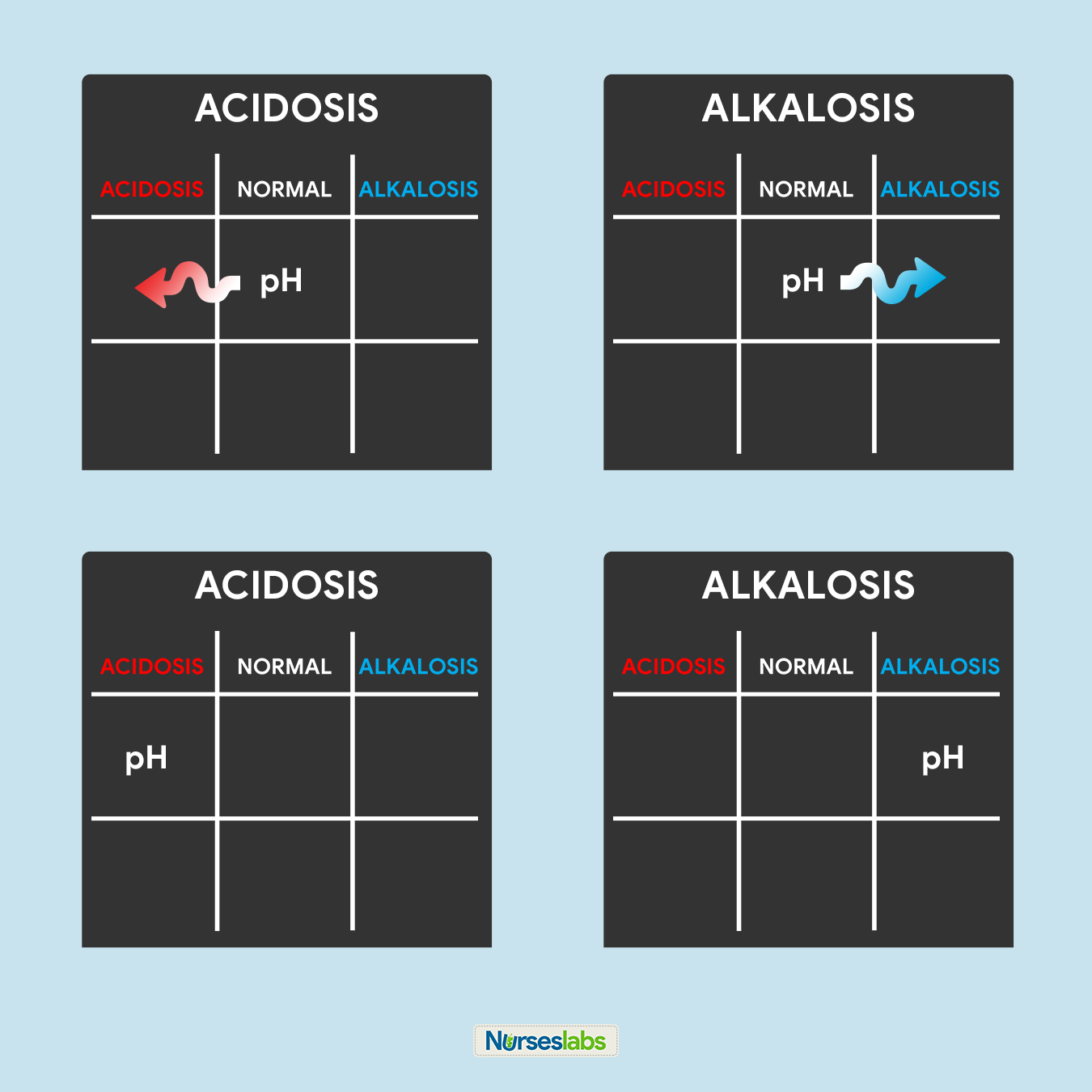
  • Unless the lost fluid and electrolytes are replaced adequately, the infected person may develop shock from profound dehydration and acidosis from loss of bicarbonate.
  • The enterotoxin acts locally and does not invade the intestinal wall. As a result, few neutrophils are found in the stool.

Causes

Cholera can be an endemic, epidemic, or a pandemic disease.

  • Environmental factors. Primary infection in humans is incidentally acquired. Risk of primary infection is facilitated by seasonal increases in the number of organisms, possibly associated with changes in water temperature and algal blooms; secondary transmission occurs through fecal-oral spread of the organism through person-to-person contact or through contaminated water and food.
  • Host factors. Malnutrition increases susceptibility to cholera. Because gastric acid can quickly render an inoculum of V cholerae noninfectious before it reaches the site of colonization in the small bowel, hydrochlorhydria or achlorhydria of any cause (including Helicobacter pylori infection, gastric surgery, vagotomy, use of H2 blockers for ulcer disease) increases susceptibility; infection rates of household contacts of cholera patients range from 20-50%. Rates are lower in areas where infection is endemic and individuals, especially adults, may have preexisting vibriocidal antibodies from previous encounters with the organism.

Statistics and Incidences

In the United States, cholera has virtually been eliminated because of improved hygiene and sanitation systems.

  • The frequency of cholera among international travelers returning to the United States has averaged 1 case per 500,000 population, with a range of 0.05-3.7 cases per 100,000 population, depending on the countries visited.
  • Between January 1, 1995, and December 31, 2000, 61 cases of cholera were reported in 18 states and 2 US territories.
  • In 1990, fewer than 30,000 cases were reported to the WHO.
  • From 2005 to 2008, 178,000-237,000 cases and 4000-6300 deaths were reported annually worldwide.
  • In nonendemic areas, the incidence of infection is similar in all age groups, although adults are less likely to become symptomatic than children.

Clinical Manifestations

After a 24- to 48-hour incubation period, symptoms begin with the sudden onset of painless watery diarrhea that may quickly become voluminous and is often followed by vomiting.

  • Diarrhea. Profuse watery diarrhea is a hallmark of cholera; cholera should be suspected when a patient older than 5 years develops severe dehydration from acute, severe, watery diarrhea (usually without vomiting) or in any patient older than 2 years who has acute watery diarrhea and is in an area where an outbreak of cholera has occurred.
  • Vomiting. Vomiting, although a prominent manifestation, may not always be present; early in the course of the disease, vomiting is caused by decreased gastric and intestinal motility; later in the course of the disease it is more likely to result from acidemia.
  • Dehydration. If untreated, the diarrhea and vomiting lead to isotonic dehydration, which can lead to acute tubular necrosis and renal failure; because the dehydration is isotonic, water loss is proportional between 3 body compartments, intracellular, intravascular, and interstitial.

Assessment and Diagnostic Findings

Definitive diagnosis is not a prerequisite for the treatment of patients with cholera.

  • Stool examination. Although observed as a gram-negative organism, the characteristic motility of Vibrio species cannot be identified on a Gram stain, but it is easily seen on direct dark-field examination of the stool.
  • Stool culture. V cholerae is not fastidious in nutritional requirements for growth; however, it does need an adequate buffering system if fermentable carbohydrate is present because viability is severely compromised if the pH is less than 6, often resulting in autosterilization of the culture.
  • Serotyping and biotyping. Specific antisera can be used in immobilization tests; a positive immobilization test result (ie, cessation of motility of the organism) is produced only if the antiserum is specific for the Vibrio type present; the second antiserum serves as a negative control.
  • Hematologic tests. Hematocrit, serum-specific gravity, and serum protein are elevated in dehydrated patients because of resulting hemoconcentration; when patients are first observed, they generally have a leukocytosis without a left shift.
  • Metabolic panel. Serum sodium is usually 130-135 mmol/L, reflecting the substantial loss of sodium in the stool; serum potassium usually is normal in the acute phase of the illness, reflecting the exchange of intracellular potassium for extracellular hydrogen ion in an effort to correct the acidosis; hyperglycemia may be present, secondary to systemic release of epinephrine, glucagon, and cortisol due to hypovolemia; patients have elevated blood urea nitrogen and creatinine levels consistent with prerenal azotemia.

Medical Management

Rehydration is the first priority in the treatment of cholera. Rehydration is accomplished in 2 phases: rehydration and maintenance.

  • Rehydration phase. The goal of the rehydration phase is to restore normal hydration status, which should take no more than 4 hours; set the rate of intravenous infusion in severely dehydrated patients at 50-100 mL/kg/hr; Lactated Ringer solution is preferred over isotonic sodium chloride solution because saline does not correct metabolic acidosis.
  • Maintenance phase. The goal of the maintenance phase is to maintain normal hydration status by replacing ongoing losses; the oral route is preferred, and the use of oral rehydration solution (ORS) at a rate of 500-1000 mL/hr is recommended.
  • Cholera cots. In areas where cholera is endemic, cholera cots have been used to assess the volume of ongoing stool losses; a cholera cot is a cot covered by a plastic sheet with a hole in the center to allow the stool to collect in a calibrated bucket underneath.
  • Diet. Resume feeding with a normal diet when vomiting has stopped; continue breastfeeding infants and young children.

Pharmacological Management

Antimicrobial therapy for cholera is an adjunct to fluid therapy and is not an essential therapeutic component.

  • Antibiotics. Empiric antimicrobial therapy must be comprehensive and should cover all likely pathogens in the context of the clinical setting; although not necessarily curative, treatment with an antibiotic to which the organism is susceptible diminishes the duration and volume of the fluid loss and hastens clearance of the organism from stool.
  • Vaccines. In June, 2016, the first U.S. cholera vaccine was approved by the FDA; contains live attenuated cholera bacteria that replicate in the gastrointestinal tract of the recipient to provide immunity; it is indicated for active immunization against disease caused by Vibrio cholerae serogroup O1 in adults aged 18-64 y traveling to cholera-affected areas.

Nursing Management

The nursing care of a client with cholera include the following:

Nursing Assessment

Assessment of the patient with cholera are as follows:

  • Assess for dehydration. Assess the status of dehydration ( skin color, temperature, skin turgor, mucous membranes, eyes, crown, body temperature, pulse, respiration, behavior, weight loss).
  • Observe for diarrhea. Observe for a sudden attack of diarrhea, fever, anorexia, vomiting, nausea, abdominal cramps, increased bowel sounds, and bowel movements more than 3 times a day, with liquid stool consistency, with or without mucus or blood.
  • Assess the level of knowledge of the family. Assess for the knowledge of diarrhea at home, dietary knowledge, and knowledge about the prevention of recurrent diarrhea.

Nursing Diagnosis

Based on the assessment data, the major nursing diagnosis for cholera are:

  • Deficient fluid volume related to excessive fluid loss through the stool or emesis.
  • Imbalanced Nutrition: less than body requirements related to loss of fluids through diarrhea, inadequate intake.
  • Risk for infection related to microorganisms that penetrate the gastrointestinal tract.
  • Impaired Skin Integrity: perianal, related to irritation from diarrhea.
  • Anxiety related to separation from parents, unfamiliar environment, a stressful procedure.

Nursing Care Planning and Goals

The major nursing care planning goals for cholera:

  • Patient will maintain adequate hydration.
  • Patient will consume adequate nutritional requirements.
  • Patient will prevent onset of infection.
  • Patient will maintain skin integrity.
  • Patient will prevent anxiety.

Nursing Interventions

The nursing interventions on a patient diagnosed with cholera are:

  • Monitor intake and output. Note number, character, and amount of stools; estimate insensible fluid losses like diaphoresis; measure urine specific gravity and observe for oliguria.
  • Weigh daily. Daily weight is an indicator of overall fluid and nutritional status.
  • Maintain hydration. Replace ongoing fluid losses until diarrhea stops.
  • Administer medications as indicated. Give an oral antibiotic to the patient with severe dehydration as prescribed.

Evaluation

Nursing goals are met as evidenced by:

  • Patient was able to maintain adequate hydration.
  • Patient was able to consume adequate nutritional requirements.
  • Patient was able to prevent onset of infection.
  • Patient was able to maintain skin integrity.
  • Patient was able to prevent anxiety.

Documentation Guidelines

Documentation in a patient with cholera include the following:

  • Individual findings, including factors affecting, interactions, nature of social exchanges, specifics of individual behavior.
  • Cultural and religious beliefs, and expectations.
  • Plan of care.
  • Teaching plan.
  • Responses to interventions, teaching, and actions performed.
  • Attainment or progress toward the desired outcome.

Practice Quiz: Cholera

Nursing practice questions for cholera. For more practice questions, visit our NCLEX practice questions page.

Exam Mode

In Exam Mode: All questions are shown but the results, answers, and rationales (if any) will only be given after you’ve finished the quiz.

Practice Quiz: Cholera

Start
Congratulations - you have completed Practice Quiz: Cholera. You scored %%SCORE%% out of %%TOTAL%%. Your performance has been rated as %%RATING%%
Your answers are highlighted below.

Practice Mode

Practice Mode: This is an interactive version of the Text Mode. All questions are given in a single page and correct answers, rationales or explanations (if any) are immediately shown after you have selected an answer. No time limit for this exam.

Practice Quiz: Cholera

Start
Congratulations - you have completed Practice Quiz: Cholera. You scored %%SCORE%% out of %%TOTAL%%. Your performance has been rated as %%RATING%%
Your answers are highlighted below.

Text Mode

Text Mode: All questions and answers are given on a single page for reading and answering at your own pace. Be sure to grab a pen and paper to write down your answers.

1. A 90-year-old client is confined to the unit for two weeks. He has been receiving antibiotics for more than a week and tells that he is having frequent watery stools. Which action will you take first?

A. Place the client on contact precautions
B. Educate the client about correct hand washing
C. Notify the physician about the loose stools
D. Get stool specimens for culture

1. Answer: A. Place the client on contact precautions.

  • Option A: Prioritization. The client may have Clostridium difficile infection based on his age, history of antibiotic therapy, and watery stools. The initial action should be to place him on contact precautions to prevent the spread of C. difficile to other clients.
  • Options B, C, and D: The other actions are also necessary and should be taken after placing the client on contact precautions.

2. A client who has frequent watery stool is admitted to the unit due to dehydration. Which nursing action should the charge nurse delegate to an LPN/LVN?

A. Giving the ordered metronidazole (Flagyl) 500 mg PO to the client
B. Reconsidering the client’s medical history for any risk factors for diarrhea
C. Doing ongoing assessments to determine the client’s hydration status
D. Explaining the purpose of ordered stool cultures to the client family

2. Answer: A. Giving the ordered metronidazole (Flagyl) 500 mg PO to the client.

  • Option A: Delegation. LPN/LVN scope of practice and education include administration of medications.
  • Options B, C, and D: Assessment of hydration status, client and family education, and assessment of client risk factors for diarrhea should be done by the RN.

3. The nurse is caring for a 20 lbs (9 kg) 6 month-old with a 3-day history of diarrhea, occasional vomiting and fever. Peripheral intravenous therapy has been initiated, with 5% dextrose in 0.33% normal saline with 20 mEq of potassium per liter infusing at 35 ml/hr. Which finding should be reported to the healthcare provider immediately?

A. 3 episodes of vomiting in 1 hour
B. Periodic crying and irritability
C. Vigorous sucking on a pacifier
D. No measurable voiding in 4 hours

3. Answer: D. No measurable voiding in 4 hours.

  • Option D: The concern is possible hyperkalemia, which could occur with continued potassium administration and a decrease in urinary output since potassium is excreted via the kidneys.

4. A 5-month old infant was brought by his mother to the health center because of diarrhea occurring 4 to 5 times a day. His skin goes back slowly after a skin pinch and his eyes are sunken. Using the IMCI guidelines, you will classify this infant in which category?

A. No signs of dehydration
B. Some dehydration
C. Severe dehydration
D. The data is insufficient

4. Answer: B. Some dehydration.

  • Option B: Using the assessment guidelines of IMCI, a child (2 months to 5 years old) with diarrhea is classified as having SOME DEHYDRATION if he shows 2 or more of the following signs: restless or irritable, sunken eyes, the skin goes back slow after a skin pinch.

5. Based on the assessment, you classified a 3-month old infant with the chief complaint of diarrhea in the category of SOME DEHYDRATION. Based on the IMCI management guidelines, which of the following will you do?

A. Bring the infant to the nearest facility where IV fluids can be given
B. Supervise the mother in giving 200 to 400 ml of Oresol in 4 hours
C. Give the infant’s mother instructions on home management
D. Keep the infant in your health center for close observation

5. Answer: B. Supervise the mother in giving 200 to 400 ml. of Oresol in 4 hours.

  • Option B: In the IMCI management guidelines, SOME DEHYDRATION is treated with the administration of Oresol within a period of 4 hours. The amount of Oresol is best computed on the basis of the child’s weight (75 ml/kg body weight). If the weight is unknown, the amount of Oresol is based on the child’s age.

References

Sources and references for this cholera study guide:

  • Black, J. M., & Hawks, J. H. (2005). Medical-surgical nursing. Elsevier Saunders,. [Link]
  • Cholera – Vibrio cholerae infection | Cholera | CDC. (2020). Retrieved 1 March 2020, from https://www.cdc.gov/cholera/
  • Willis, L. (2019). Professional guide to diseases. Lippincott Williams & Wilkins. [Link]

Middle East Respiratory Syndrome Coronavirus (MERS-CoV)

$
0
0

Review this study guide to know more about the Middle East respiratory syndrome, its causes, symptoms, treatment, prevention, and nursing management.

MERS or Middle East respiratory syndrome is a zoonotic disease (spreads from animals to people) that can cause severe respiratory illness. It was first identified in Saudi Arabia in 2012 and has infected more than 2,000 individuals worldwide.

What is Middle East Respiratory Syndrome (MERS)?

Middle East respiratory syndrome (MERS) is caused by a novel coronavirus (Middle East respiratory syndrome coronavirus, or MERS‐CoV).

  • Through first reported in Saudi Arabia, it was later identified that the first known cases of MERS occurred in Jordan in April 2012.
  • Most MERS patients developed severe respiratory illness with symptoms of fever, cough, and shortness of breath.
  • A large MERS outbreak occurred in the Republic of South Korea linked to a traveler from the Arabian Peninsula in 2015.
  • Travel-associated cases have been identified in Algeria, Austria, China, Egypt, France, Germany, Greece, Italy, Malaysia, Netherlands, Philippines, Republic of Korea, Thailand, Tunisia, Turkey, United Kingdom (UK), and United States (US).
  • CDC has published guidance for health departments and healthcare infection-control programs for investigating potential cases of MERS and preventing its spread.

Pathophysiology

MERS is considered an international threat to public health.

  • Compared with severe acute respiratory syndrome coronavirus (SARS-Cov), MERS-CoV can establish infection in monocyte-derived macrophages (MDMs) and macrophages.
  • The virus induces the release of proinflammatory cytokines, leading to severe inflammation and tissue damage, which may manifest clinically as severe pneumonia and respiratory failure. [
  • Vascular endothelial cells located in the pulmonary interstitium may also be infected by MERS-CoV, and, because MERS-CoV receptor DPP4 is expressed in different human cells and tissues, dissemination of the infection may occur. 
  • Interestingly, lymphopenia has been noted in most patients infected with MERS-CoV, as was noted in SARS infections.
  • This is due to cytokine-induced immune cell sequestration and release and induction of monocyte chemotactic protein-1 (MCP-1) and interferon-gamma-inducible protein-10 (IP-10), which suppresses the proliferation of human myeloid progenitor cells.

Causes

Coronaviruses are the largest of all RNA viruses, with positive-sense single-stranded RNA genomes of 26-32 kb.

  • Betacoronavirus. MERS-CoV is a recently discovered betacoronavirus of lineage C that was first reported in Saudi Arabia in 2012; the exact origin of this novel coronavirus is still unknown; MERS-CoV is closely related to two coronaviruses of the same lineage found in bats, which may indeed be its wild reservoir.
  • Dromedary camels. Specific mechanisms for transmission from animals are unclear but appear to involve contact with dromedary camels or their urine, as well as the consumption of their undercooked meat or unpasteurized dairy products.

Statistics and Incidences

About 3 or 4 out of every 10 patients reported with MERS have died.

  • In May 2014, CDC confirmed two unlinked imported cases of MERS in the United States—one to Indiana, the other to Florida; both cases were among healthcare providers who lived and worked in Saudi Arabia; both traveled to the U.S. from Saudi Arabia, where scientists believe they were infected.
  • Since 2012, 2,374 laboratory-confirmed cases of infection with MERS-CoV have been reported to the World Health Organization (WHO), including at least 823 related deaths.
  • Twenty-seven countries have reported MERS cases.
  • On the Arabian Peninsula, countries include Bahrain, Iran, Jordan, Kuwait, Lebanon, Oman, Qatar, Saudi Arabia, United Arab Emirates (UAE), and Yemen.
  • Other countries reporting travel-associated MERS include Algeria, Austria, China, Egypt, France, Germany, Greece, Italy, Malaysia, Netherlands, Philippines, Republic of Korea, Thailand, Tunisia, Turkey, United Kingdom (UK), and the United States.
  • The vast majority of these cases have so far occurred in the Kingdom of Saudi Arabia.
  • The largest MERS outbreak outside of Saudi Arabia occurred in 2015 in the Republic of Korea; the outbreak involved 186 confirmed cases and caused 36 deaths.
  • The outbreak sparked quarantine of more than 5,000 individuals and the closure of 2,000 schools before ending.

Clinical Manifestations

Physical examination findings associated with MERS-CoV infection are similar to those presenting with any flu-like symptoms, including the following:

  • Fever
  • Rhinorrhea, mostly clear
  • Pulmonary findings, including hypoxemia, rhonchi, and rales (some patients may have a normal auscultation)
  • Tachycardia
  • Hypotension may occur with severe illness, reflecting systemic inflammatory response syndrome

Assessment and Diagnostic Findings

Most state laboratories are approved to test for Middle East Respiratory Syndrome Coronavirus (MERS-CoV) using CDC’s rRT-PCR assay.

  • rRT-PCR assay. FDA issued an Emergency Use Authorization (EUA) on June 5, 2013, to authorize use of CDC’s 2012 real-time reverse transcription–PCR assay to test for MERS-CoV in clinical respiratory, serum, and stool specimens.
  • Serology. Serologic testing for MERS-CoV is available as a research/surveillance test from the CDC; it is not considered a diagnostic test but may offer valuable epidemiologic data; it must be ordered in consultation and with approval of CDC via the EOC.
  • Laboratory studies. Laboratory findings at presentation may include leukopenia, lymphopenia, thrombocytopenia, and elevated lactate dehydrogenase levels; these are most likely with increasing severity of illness. 
  • Imaging studies. Chest imaging findings are abnormal in more than 80% of MERS cases; ground-glass opacity (GGO) is found in over 60% of chest radiographs, with about 20% incidence of consolidation; some infiltrates may be nodular.

Medical Management

Management of the Middle East respiratory syndrome (MERS) coronavirus (MERS-CoV) infection is supportive; this includes hydration, antipyretic, analgesics, respiratory support, and antibiotics if needed for bacterial superinfection.

  • Consultations. Upon suspicion of MERS, the patient should be placed in an airborne infection isolation room (AIIR) with a minimum of 12 air exchanges per hour, and personnel protection equipment (PEP) appropriate for contact and airborne precautions (gown, gloves, goggles, and N-95 respirator mask or powered air purifier respirator [PAPR]) should be used.
  • Medical care. Medical care is supportive and depends on the severity of illness.
  • Prevention. No MERS-CoV vaccine is commercially available; prevention of infection in areas where MERS-CoV is being actively transmitted requires avoidance of potentially infectious secretions and careful attention to hand and respiratory hygiene.

Pharmacologic Management

No medications have been approved for the treatment of coronavirus infections. Clinical trials are needed to establish any benefit from ribavirin and/or interferon alfa.

Nursing Management

Nursing care for a patient with MERS-CoV include the following:

Nursing Assessment

Assessment of a patient with MERS-CoV include:

  • History. A high index of suspicion is necessary to suspect MERS, and a travel and exposure history is essential to the diagnosis; keys to the case definition of MERS is a history of residence or travel in the Arabian Peninsula, in countries where MERS-CoV is known to be circulating in dromedary camels, or where human infections have recently occurred and exposure within the incubation period of 14 days.
  • Physical exam. Clinical manifestation is indistinguishable from other common respiratory viruses and may range from no symptoms to rapidly progressive multiorgan failure and death.

Nursing Diagnosis

Based on the assessment data, the major nursing diagnosis for a patient with MERS-CoV include the following:

  • Infection related to failure to avoid pathogen secondary to exposure to MERS-CoV.
  • Deficient knowledge related to unfamiliarity with disease transmission information.
  • Hyperthermia related to increase in metabolic rate.
  • Ineffective airway clearance related to excessive production of pulmonary secretions.
  • Anxiety related to unknown etiology of the disease.

Nursing Care Planning and Goals

The major nursing care plan goals for a patient with MERS-CoV are:

  • Prevent the spread of infection.
  • Learn more about the disease and its management.
  • Reduce increase in temperature.
  • Provide a patent airway.
  • Reduce anxiety.

Nursing Interventions

Nursing interventions for the patient with MERS-CoV include the following:

  • Monitor vital signs. Monitor the patient’s temperature; the infection usually begins with a high temperature; monitor the respiratory rate of the patient as shortness of breath is another common symptom.
  • Educate the patient and folks. Include the patient and folks in creating the teaching plan, beginning with establishing objectives and goals for learning at the beginning of the session; provide clear, thorough, and understandable explanations and demonstrations; and give information with the use of media. Use visual aids like diagrams, pictures, videotapes, audiotapes, and interactive Internet websites, such as Nurseslabs.
  • Reduce increase in temperature. Adjust and monitor environmental factors like room temperature and bed linens as indicated; encourage ample fluid intake by mouth; eliminate excess clothing and covers, and give antipyretic medications as prescribed.
  • Ensure patent airway. Teach the patient the proper ways of coughing and breathing. (e.g., take a deep breath, hold for 2 seconds, and cough two or three times in succession); position the patient upright if tolerated, and encourage patient to increase fluid intake to 3 liters per day within the limits of cardiac reserve and renal function.
  • Reduce anxiety. Use presence, touch (with permission), verbalization, and demeanor to remind patients that they are not alone and to encourage expression or clarification of needs, concerns, unknowns, and questions; accept patient’s defenses; do not dare, argue, or debate; converse using a simple language and brief statements; and allow the patient to talk about anxious feelings and examine anxiety-provoking situations if they are identifiable.

Evaluation

Nursing evaluation of goals for a patient with MERS-COV are met as evidenced by:

  • Prevention of the spread of infection.
  • Acquired knowledge about the disease and its management.
  • Reduction in levels of temperature.
  • Patent airway achieved.
  • Reduction in anxiety.

Documentation Guidelines

Documentation guidelines for a patient with MERS-CoV include the following:

  • Individual findings, including factors affecting, interactions, nature of social exchanges, specifics of individual behavior.
  • Cultural and religious beliefs, and expectations.
  • Plan of care.
  • Teaching plan.
  • Responses to interventions, teaching, and actions performed.
  • Attainment or progress toward the desired outcome.

Practice Quiz: Middle East Respiratory Syndrome (MERS)

Nursing practice questions for Middle East Respiratory Syndrome (MERS). For more practice questions, visit our NCLEX practice questions page.

Exam Mode

In Exam Mode: All questions are shown but the results, answers, and rationales (if any) will only be given after you’ve finished the quiz.

Practice Quiz: Middle East Respiratory Syndrome (MERS)

Start

Congratulations - you have completed Practice Quiz: Middle East Respiratory Syndrome (MERS).

You scored %%SCORE%% out of %%TOTAL%%.

Your performance has been rated as %%RATING%%


Your answers are highlighted below.

Practice Mode

Practice Mode: This is an interactive version of the Text Mode. All questions are given in a single page and correct answers, rationales or explanations (if any) are immediately shown after you have selected an answer. No time limit for this exam.

Practice Quiz: Middle East Respiratory Syndrome (MERS)

Start

Congratulations - you have completed Practice Quiz: Middle East Respiratory Syndrome (MERS).

You scored %%SCORE%% out of %%TOTAL%%.

Your performance has been rated as %%RATING%%


Your answers are highlighted below.

Text Mode

Text Mode: All questions and answers are given on a single page for reading and answering at your own pace. Be sure to grab a pen and paper to write down your answers.

1. In which order will the nurse perform the following actions as she prepares to leave the room of a client with airborne precautions after performing oral suctioning?

1. Take off goggles
2. Take off gown
3. Remove gloves
4. Remove N95 respirator
5. Perform hand hygiene

A. 1, 2, 3, 4, 5
B. 2, 1, 4, 3, 5
C. 3, 4, 1, 2, 5
D. 4, 3, 2, 1, 5

1. Answer: B. 2, 1, 4, 3, 5

  • Option B: Prioritization. This order will prevent contact of the contaminated gloves and gown with areas like the hair that cannot be readily cleaned after client contact and stop transmission of microorganisms to the nurse and the client. The correct order for donning and removal of PPE has been standardized by agencies such as the Centers for Disease Control and Prevention (CDC) and the Occupational Safety and Health Administration.

2. The nurse assessed the client and noted shortness of breath and recent trip to China. The client is strongly suspected of having Middle East Respiratory Syndrome (MERS-CoV). Which of these prescribed actions will the nurse take first?

A. Place the client on airborne and contact precautions
B. Introduce normal saline at 75 mL/hr
C. Give methylprednisolone (SOLU-Medrol) 1 g intravenously (IV)
D. Take blood, urine, sputum cultures

2. Answer: A. Place the client on airborne and contact precautions

  • Option A: Prioritization. MERS-CoV is considered deadly so the initial action is to protect other clients and healthcare workers by securing the client in isolation. If an airborne-agent isolation (negative-pressure) room is not yet available, droplet precautions should be initiated until the client can be moved to a negative-pressure room.
  • Options B, C, and D: The other actions should also be taken immediately but are not as important as preventing transmission of the disease.

3. The school nurse is asked which action will take to have the most impact on the incidence of infectious diseases in school. The correct response is:

A. Grant written information about infection control to all parents
B. Ensure that students are immunized according to national guidelines
C. Make soap and water easily accessible in the classrooms
D. Educate students on how to cover their mouths when coughing

3. Answer: B. Ensure that students are immunized according to national guidelines.

  • Option B: Prioritization. The incidence of once-common infectious diseases like measles, chickenpox, and mumps has been most effectively reduced by immunization of all school-aged children.
  • Options A, C, and D: The other options are also important but will not have much impact as immunization.

4. Which of the following is the FIRST priority in preventing infections when providing care for a client?

A. Handwashing
B. Wearing gloves
C. Using a barrier between client’s furniture and nurse’s bag
D. Wearing gowns and goggles

4. Answer: A. Handwashing

  • Option A: Handwashing remains the most effective way to avoid spreading infection. However, too often nurses do not practice good handwashing techniques and do not teach families to do so. Nurses need to wash their hands before and after touching the client and before entering the nursing bag.

5. A 10-year-old client contracted Middle East Respiratory Syndrome (MERS-CoV) when traveling abroad with her parents. The nurse knows she must put on personal protective equipment to protect herself while providing care. Based on the mode of MERS-CoV transmission, which personal protective should the nurse wear?

A. Gloves
B. Gown and gloves
C. Gown, gloves, and mask
D. Gown, gloves, mask, and eye goggles or eye shield

5. Answer D. Gown, gloves, mask, and eye goggles or eye shield

  • Option D: The transmission of MERS-CoV isn’t fully understood. Therefore, all modes of transmission must be considered possible, including airborne, droplet, and direct contact with the virus. For protection from contracting MERS-CoV, any health care worker providing care for a client with MERS should wear a gown, gloves, mask, and eye goggles or an eye shield.

References

Sources and references for this study guide for MERS-COV:

  • de Groot, R. J., Baker, S. C., Baric, R. S., Brown, C. S., Drosten, C., Enjuanes, L., … & Perlman, S. (2013). Commentary: Middle East respiratory syndrome coronavirus (MERS-CoV): announcement of the Coronavirus Study Group. Journal of virology87(14), 7790-7792. [Link]
  • Kimberlin, D. W. (2018). Red Book: 2018-2021 report of the committee on infectious diseases (No. Ed. 31). American academy of pediatrics.
  • Oshinsky, D. M. (2005). Polio: an American story. Oxford University Press. [Link]
  • Willis, L. (2019). Professional guide to diseases. Lippincott Williams & Wilkins. [Link]

IV Fluids and Solutions Guide & Cheat Sheet (2020 Update)

$
0
0

Get to know the different types of intravenous solutions or IV fluids in this guide and cheat sheet. Differentiate isotonic, hypertonic, and hypotonic IV solutions and the nursing interventions and management for each. 

What are IV Fluids? 

Intravenous fluids, also known as intravenous solutions, are supplemental fluids used in intravenous therapy to restore or maintain normal fluid volume and electrolyte balance when the oral route is not possible. IV fluid therapy is an efficient and effective way of supplying fluids directly into the intravascular fluid compartment, in replacing electrolyte losses, and in administering medications and blood products. 

Types of IV Fluids

There are different types of IV fluids and different ways on how to classify them.

The most common way to categorize IV fluids is based on their tonicity:

  • Isotonic. Isotonic IV solutions that have the same concentration of solutes as blood plasma.
  • Hypotonic. Hypotonic solutions have lesser concentration of solutes than plasma.
  • Hypertonic. Hypertonic solutions have greater concentration of solutes than plasma.

IV solutions can also be classified based on their purpose:

  • Nutrient solutions. May contain dextrose, glucose, and levulose to make up the carbohydrate component – and water. Water is supplied for fluid requirements and carbohydrate for calories and energy. Nutrient solutions are useful in preventing dehydration and ketosis. Examples of nutrient solutions include D5W, D5NSS.
  • Electrolyte solutions. Contains varying amounts of cations and anions that are used to replace fluid and electrolytes for clients with continuing losses. Examples of electrolyte solutions include 0.9 NaCl, Ringer’s Solution, and LRS.
  • Alkalinizing solutions. Are administered to treat metabolic acidosis. Examples: LRS.
  • Acidifying solutions. Are used to counteract metabolic alkalosis. D51/2NS, 0.9 NaCl.
  • Volume expanders. Are solutions used to increase the blood volume after a severe blood loss, or loss of plasma. Examples of volume expanders are dextran, human albumin, and plasma.

Crystalloids

Crystalloid IV solutions contain small molecules that flow easily across semipermeable membranes. They are categorized according to their relative tonicity in relation to plasma. There are three types: isotonic, hypotonic, and hypertonic.

Isotonic IV Fluids

Isotonic IV Fluids Cheat Sheet
Cheat sheet for Isotonic IV Fluids.

Most IV fluids are isotonic, meaning, they have the same concentration of solutes as blood plasma. When infused, isotonic solutions expand both the intracellular fluid and extracellular fluid spaces, equally. Such fluids do not alter the osmolality of the vascular compartment. Technically, electrolyte solutions are considered isotonic if the total electrolyte content is approximately 310 mEq/L. Isotonic IV fluids have a total osmolality close to that of the ECF and do not cause red blood cells to shrink or swell.

0.9% NaCl (Normal Saline Solution, NSS)

Normal saline solution (0.9% NaCl) or NSS, is a crystalloid isotonic IV fluid that contains water, sodium (154 mEq/L), and chloride (154 mEq/L). It has an osmolality of 308 mOsm/L and gives no calories. It is called normal saline solution because the percentage of sodium chloride dissolved in the solution is similar to the usual concentration of sodium and chloride in the intravascular space. Normal saline is the isotonic solution of choice for expanding the extracellular fluid (ECF) volume because it does not enter the intracellular fluid (ICF). It is administered to correct extracellular fluid volume deficit because it remains within the ECF. 

Normal saline is the IV fluid used alongside the administration of blood products. It is also used to replace large sodium losses such as in burn injuries and trauma. It should not be used for heart failure, pulmonary edema, and renal impairment, or conditions that cause sodium retention as it may risk fluid volume overload. 

Dextrose 5% in Water (D5W)

D5W (dextrose 5% in water) is a crystalloid isotonic IV fluid with a serum osmolality of 252 mOsm/L. D5W is initially an isotonic solution and provides free water when dextrose is metabolized (making it a hypotonic solution), expanding the ECF and the ICF. It is administered to supply water and to correct an increase in serum osmolality. A liter of D5W provides fewer than 200 kcal and contains 50g of glucose. It should not be used for fluid resuscitation because hyperglycemia can result. It should also be avoided to be used in clients at risk for increased intracranial pressure as it can cause cerebral edema. 

Lactated Ringer’s 5% Dextrose in Water (D5LRS) 

Lactated Ringer’s Solution (also known as Ringer’s Lactate or Hartmann solution) is a crystalloid isotonic IV fluid designed to be the near-physiological solution of balanced electrolytes. It contains 130 mEq/L of sodium, 4 mEq/L of potassium, 3 mEq/L of calcium, and 109 mEq/L of chloride. It also contains bicarbonate precursors to prevent acidosis. It does not provide calories or magnesium and has limited potassium replacement. It is the most physiologically adaptable fluid because its electrolyte content is most closely related to the composition of the body’s blood serum and plasma. 

Lactated Ringer’s is used to correct dehydration, sodium depletion, and replace GI tract fluid losses. It can also be used in fluid losses due to burns, fistula drainage, and trauma. It is the choice for first-line fluid resuscitation for certain patients. It is often administered to patients with metabolic acidosis. 

Lactated Ringer’s solution is metabolized in the liver, which converts the lactate to bicarbonate, therefore, it should not be given to patients who cannot metabolize lactate (e.g., liver disease, lactic acidosis). It should be used in caution for patients with heart failure and renal failure. 

Ringer’s Solution

Ringer’s solution is another isotonic IV solution that has content similar to Lactated Ringer’s Solution but does not contain lactate. Indications are the same for Lactated Ringer’s but without the contraindications related to lactate. 

Nursing Considerations for Isotonic Solutions

The following are the general nursing interventions and considerations when administering isotonic solutions:

  • Document baseline data. Before infusion, assess the patient’s vital signs, edema status, lung sounds, and heart sounds. Continue monitoring during and after the infusion. 
  • Observe for signs of fluid overload. Look for signs of hypervolemia such as hypertension, bounding pulse, pulmonary crackles, dyspnea, shortness of breath, peripheral edema, jugular venous distention, and extra heart sounds. 
  • Monitor manifestations of continued hypovolemia. Look for signs that indicate continued hypovolemia such as, decreased urine output, poor skin turgor, tachycardia, weak pulse, and hypotension.
  • Prevent hypervolemia. Patients being treated for hypovolemia can quickly develop fluid overload following rapid or over infusion of isotonic IV fluids. 
  • Elevate the head of the bed at 35 to 45 degrees. Unless contraindicated, position the client in semi-Fowler’s position. 
  • Elevate the patient’s legs. If edema is present, elevate the legs of the patient to promote venous return.
  • Educate patients and families. Teach patients and families to recognize signs and symptoms of fluid volume overload. Instruct patients to notify their nurse if they have trouble breathing or notice any swelling. 
  • Close monitoring for patients with heart failure. Because isotonic fluids expand the intravascular space, patients with hypertension and heart failure should be carefully monitored for signs of fluid overload. 

Hypotonic IV Fluids

Hypotonic IV solutions have a lower osmolality and contain fewer solutes than plasma. They cause fluid shifts from the ECF into the ICF to achieve homeostasis, therefore, causing cells to swell and may even rupture. IV solutions are considered hypotonic if the total electrolyte content is less than 250 mEq/L. Hypotonic IV fluids are usually used to provide free water for excretion of body wastes, treat cellular dehydration, and replace the cellular fluid. 

0.45% Sodium Chloride (0.45% NaCl)

Sodium chloride 0.45% (1/2 NS), also known as half-strength normal saline, is a hypotonic IV solution used for replacing water in patients who have hypovolemia with hypernatremia. Excess use may lead to hyponatremia due to the dilution of sodium, especially in patients who are prone to water retention. It has an osmolality of 154 mOsm/L and contains 77 mEq/L sodium and chloride. Hypotonic sodium solutions are used to treat hypernatremia and other hyperosmolar conditions. 

0.33% Sodium Chloride (0.33% NaCl)

Hypotonic IV Fluids and Solutions Cheat Sheet
Cheat sheet for Hypotonic IV Fluids.

0.33% Sodium Chloride Solution is used to allow kidneys to retain the needed amounts of water and is typically administered with dextrose to increase tonicity. It should be used in caution for patients with heart failure and renal insufficiency. 

0.225% Sodium Chloride (0.225% NaCl)

0.225% Sodium Chloride Solution is often used as a maintenance fluid for pediatric patients as it is the most hypotonic IV fluid available at 77 mOsm/L. Used together with dextrose. 

2.5% Dextrose in Water (D2.5W)

Another hypotonic IV solution commonly used is 2.5% dextrose in water (D2.5W). This solution is used to treat dehydration and decreased the levels of sodium and potassium. It should not be administered with blood products as it can cause hemolysis of red blood cells. 

Nursing Considerations for Hypotonic Solutions

The following are the general nursing interventions and considerations when administering hypotonic IV solutions:

  • Document baseline data. Before infusion, assess the patient’s vital signs, edema status, lung sounds, and heart sounds. Continue monitoring during and after the infusion. 
  • Do not administer in contraindicated conditions. Hypotonic solutions may exacerbate existing hypovolemia and hypotension causing cardiovascular collapse. Avoid use in patients with liver disease, trauma, or burns. 
  • Risk for increased intracranial pressure (IICP). Should not be given to patients with risk for IICP as the fluid shift may cause cerebral edema (remember: hypotonic solutions make cells swell). 
  • Monitor for manifestations of fluid volume deficit. Signs and symptoms include confusion in older adults. Instruct patients to inform the nurse if they feel dizzy. 
  • Warning on excessive infusion. Excessive infusion of hypotonic IV fluids can lead to intravascular fluid depletion, decreased blood pressure, cellular edema, and cell damage. 
  • Do not administer along with blood products. Most hypotonic solutions can cause hemolysis of red blood cells especially during rapid infusion of the solution. 

Hypertonic IV Fluids

Hypertonic IV solutions have a greater concentration of solutes (375 mEq/L and greater) than plasma and cause fluids to move out of the cells and into the ECF in order to normalize the concentration of particles between two compartments. This effect causes cells to shrink and may disrupt their function. They are also known as volume expanders as they draw water out of the intracellular space, increasing extracellular fluid volume. 

Hypertonic IV Fluids and Solutions Cheat Sheet
Cheat sheet for Hypertonic IV Fluids.

Hypertonic Sodium Chloride IV Fluids

Hypertonic sodium chloride solutions contain a higher concentration of sodium and chloride than normally contained in plasma. Infusion of hypertonic sodium chloride solution shifts fluids from the intracellular space into the intravascular and interstitial spaces. Hypertonic sodium chloride IV solutions are available in the following forms and strengths: 

  • 3% sodium chloride (3% NaCl) containing 513 mEq/L of sodium and chloride with an osmolality of 1030 mOsm/L. 
  • 5% sodium chloride (5% NaCl) containing 855 mEq/L of sodium and chloride with an osmolality of 1710 mOsm/L. 

Hypertonic sodium chloride solutions are used in the acute treatment of sodium deficiency (severe hyponatremia) and should be used only in critical situations to treat hyponatremia. They need to be infused at a very low rate to avoid the risk of overload and pulmonary edema. If administered in large quantities and rapidly, they may cause an extracellular volume excess and precipitate circulatory overload and dehydration. Therefore, they should be administered cautiously and usually only when the serum osmolality has decreased to critically low levels.  Some patients may need diuretic therapy to assist in fluid excretion. It is also used in patients with cerebral edema. 

Hypertonic Dextrose Solutions

Isotonic solutions that contain 5% dextrose (e.g., D5NSS, D5LRS) are slightly hypertonic since they exceed the total osmolality of the ECF. However, dextrose is quickly metabolized and only the isotonic solution remains. Therefore, any effect on the ICF is temporary. Hypertonic dextrose solutions are used to provide kilocalories for the patient in the short term. Higher concentrations of dextrose (i.e., D50W) are strong hypertonic solutions and must be administered into central veins so that they can be diluted by rapid blood flow. 

Dextrose 10% in Water (D10W)

Dextrose 10% in Water (D10W) is an hypertonic IV solution used in the treatment of ketosis of starvation and provides calories (380 kcal/L), free water, and no electrolytes. It should be administered using a central line if possible and should not be infused using the same line as blood products as it can cause RBC hemolysis. 

Dextrose 20% in Water (D20W) 

Dextrose 20% in Water (D20W) is hypertonic IV solution an osmotic diuretic that causes fluid shifts between various compartments to promote diuresis. 

Dextrose 50% in Water (D50W)

Another hypertonic IV solution used commonly is Dextrose 50% in Water (D50W) which is used to treat severe hypoglycemia and is administered rapidly via IV bolus. 

Nursing Considerations for Hypertonic Solutions

The following are the general nursing interventions and considerations when administering hypertonic IV solutions:

  • Document baseline data. Before infusion, assess the patient’s vital signs, edema status, lung sounds, and heart sounds. Continue monitoring during and after the infusion. 
  • Watch for signs of hypervolemia. Since hypertonic solutions move fluid from the ICF to the ECF, they increase the extracellular fluid volume and increases the risk for hypervolemia. Look for signs of swelling in arms, legs, face, shortness of breath, high blood pressure, and discomfort in the body (e.g., headache, cramping). 
  • Monitor and observe the patient during administration. Hypertonic solutions should be administered only in high acuity areas with constant nursing surveillance for potential complications. 
  • Verify order. Prescription for hypertonic solutions should state the specific hypertonic fluid to be infused, the total volume to be infused, the infusion rate and the length of time to continue the infusion. 
  • Assess health history. Patients with kidney or heart disease and those who are dehydrated should not receive hypertonic IV fluids. These solutions can affect renal filtration mechanisms and can easily cause hypervolemia to patients with renal or heart problems. 
  • Prevent fluid overload. Ensure that administration of hypertonic fluids does not precipitate fluid volume excess or overload. 
  • Do not administer peripherally. Hypertonic solutions can cause irritation and damage to the blood vessel and should be administered through a central vascular access device inserted into a central vein. 
  • Monitor blood glucose closely. Rapid infusion of hypertonic dextrose solutions can cause hyperglycemia. Use with caution for patients with diabetes mellitus. 

Colloids

Colloids contain large molecules that do not pass through semipermeable membranes. Colloids are IV fluids that contain solutes of high molecular weight, technically, they are hypertonic solutions, which when infused, exert an osmotic pull of fluids from interstitial and extracellular spaces. They are useful for expanding the intravascular volume and raising blood pressure. Colloids are indicated for patients in malnourished states and patients who cannot tolerate large infusions of fluid. 

Colloid IV Fluids and Solutions Cheat Sheet

Human Albumin

Human albumin is a solution derived from plasma. It has two strengths: 5% albumin and 25% albumin. 5% Albumin is a solution derived from plasma and is a commonly utilized colloid solution. It is used to increase the circulating volume and restore protein levels in conditions such as burns, pancreatitis, and plasma loss through trauma. 25% Albumin is used together with sodium and water restriction to reduce excessive edema. They are considered blood transfusion products and uses the same protocols and nursing precautions when administering albumin. 

The use of albumin is contraindicated in patients with the following conditions: severe anemia, heart failure, or known sensitivity to albumin. Additionally, angiotensin-converting enzyme inhibitors should be withheld for at least 24 hours before administering albumin because of the risk of atypical reactions, such as hypotension and flushing. 

Dextrans

Dextrans are polysaccharides that act as colloids. They are available in two types: low-molecular-weight dextrans (LMWD) and high-molecular-weight dextrans (HMWD). They are available in either saline or glucose solutions. Dextran interferes with blood crossmatching, so draw the patient’s blood before administering dextran, if crossmatching is anticipated. 

Low-molecular-weight Dextrans (LMWD)

LMWD contains polysaccharide molecules that behave like colloids with an average molecular weight of 40,000 (Dextran 40). LMWD is used to improve the microcirculation in patients with poor peripheral circulation. They contain no electrolytes and are used to treat shock related to vascular volume loss (e.g., burns, hemorrhage, trauma, or surgery). On certain surgical procedures, LMWDs are used to prevent venous thromboembolism. They are contraindicated in patients with thrombocytopenia, hypofibrinogenemia, and hypersensitivity to dextran. 

High-molecular-weight Dextrans (HMWD)

HMWD contains polysaccharide molecules with an average molecular weight of 70,000 (Dextran 70) or 75,000 (Dextran 75). HMWD used for patients with hypovolemia and hypotension. They are contraindicated in patients with hemorrhagic shock. 

Etherified Starch

These solutions are derived from starch and are used to increase intravascular fluid but can interfere with normal coagulation. Examples include EloHAES, HyperHAES, and Voluven. 

Gelatin 

Gelatins have lower molecular weight than dextrans and therefore remain in the circulation for a shorter period of time. 

Plasma Protein Fraction (PPF)

Plasma Protein Fraction is a solution that is also prepared from plasma, and like albumin, is heated before infusion. It is recommended to infuse slowly to increase circulating volume. 

Nursing Considerations for Colloid Solutions

The following are the general nursing interventions and considerations when administering colloid IV solutions:

  • Assess allergy history. Most colloids can cause allergic reactions, although rare, so take a careful allergy history, asking specifically if they’ve ever had a reaction to an IV infusion before. 
  • Use a large-bore needle (18-gauge). A larger needle is needed when administering colloid solutions. 
  • Document baseline data. Before infusion, assess the patient’s vital signs, edema status, lung sounds, and heart sounds. Continue monitoring during and after the infusion. 
  • Monitor the patient’s response. Monitor intake and output closely for signs of hypervolemia, hypertension, dyspnea, crackles in the lungs, and edema. 
  • Monitor coagulation indexes. Colloid solutions can interfere with platelet function and increase bleeding times, so monitor the patient’s coagulation indexes.

Cheat Sheet for IV Fluids

In this section is where you can download the cheat sheets for intravenous solutions. Simply click on the images below to open the enlarged image. Please feel free to print, share, and use in your presentations or reports.

References and Sources

The following are the references and sources for this IV fluid guide that you may find interesting or if you want to further your reading:

  • Berman, A., Snyder, S. J., Levett-Jones, T., Dwyer, T., Hales, M., Harvey, N., … & Stanley, D. (2018). Kozier and Erb’s Fundamentals of Nursing [4th Australian edition].
  • Dougherty, L., & Lamb, J. (Eds.). (2009). Intravenous therapy in nursing practice. John W
  • Hinkle, J. L., & Cheever, K. H. (2017). Brunner-Suddarth. Medical-surgical nursing.

Arterial Blood Gas Analysis Made Easy with Tic-Tac-Toe Method

$
0
0

Interpretation of arterial blood gases (ABGs) is a crucial skill that a lot of student nurses and medical practitioners need to learn. In this guide, we’ll help you understand the concepts behind arterial blood gas and teach you the easiest and most fun way to interpret ABGs using the tic-tac-toe method.

What is arterial blood gas? 

Collection of arterial blood for arterial blood gas (ABG) test.

An arterial blood gas is a laboratory test to monitor the patient’s acid-base balance. It is used to determine the extent of the compensation by the buffer system and includes the measurements of the acidity (pH), levels of oxygen, and carbon dioxide in arterial blood. Unlike other blood samples obtained through a vein, a blood sample from an arterial blood gas (ABG) is taken from an artery (commonly on radial or brachial artery). 

What are the components of arterial blood gas? 

There are six components of arterial blood gas (ABGs):

pH

The pH is the concentration of hydrogen ions and determines the acidity or alkalinity of body fluids. A pH of 7.35 indicates acidosis and a pH greater than 7.45 indicates alkalosis. The normal ABG level for pH is 7.35 to 7.45.

PaCO2 (Partial Pressure of Carbon Dioxide)

PaCO2 or partial pressure of carbon dioxide shows the adequacy of the gas exchange between the alveoli and the external environment (alveolar ventilation). Carbon dioxide (CO2) cannot escape when there is damage in the alveoli, excess CO2 combines with water to form carbonic acid (H2CO3) causing an acidotic state. When there is hypoventilation in the alveolar level (for example, in COPD), the PaCO2 is elevated, and respiratory acidosis results. On the other hand, when there is alveolar hyperventilation (e.g., hyperventilation), the PaCO2 is decreased causing respiratory alkalosis. For PaCO2, the normal range is 35 to 45 mmHg (respiratory determinant).

PaO2 (Partial Pressure of Oxygen)

PaO2 or partial pressure of oxygen or PAO2 indicates the amount of oxygen available to bind with hemoglobin. The pH plays a role in the combining power of oxygen with hemoglobin: a low pH means there is less oxygen in the hemoglobin. For PaO2, the normal range is 75 to 100 mmHg

SO2 (Oxygen Saturation)

SO2 or oxygen saturation, measured in percentage, is the amount of oxygen in the blood that combines with hemoglobin. It can be measured indirectly by calculating the PAO2 and pH Or measured directly by co-oximetry. Oxygen saturation, the normal range is 94–100%

HCO3 (Bicarbonate)

HCO3 or bicarbonate ion is an alkaline substance that comprises over half of the total buffer base in the blood. A deficit of bicarbonate and other bases indicates metabolic acidosis. Alternatively, when there is an increase in bicarbonates present, then metabolic alkalosis results. 

BE (Base Excess)

BE. Base excess or BE value is routinely checked with HCO3 value. A base excess of less than –2 is acidosis and greater than +2 is alkalosis. Base excess, the normal range is –2 to +2 mmol/L

Normal Values in Arterial Blood Gas 

To determine acid-base imbalance, you need to know and memorize these values to recognize what deviates from normal. The normal range for ABGs is used as a guide, and the determination of disorders is often based on blood pH. If the blood is basic, the HCO3 level is considered because the kidneys regulate bicarbonate ion levels. If the blood is acidic, the PaCO2 or partial pressure of carbon dioxide in arterial blood is assessed because the lungs regulate the majority of acid. The normal ABG values are the following:

  • For  pH, the normal range is 7.35 to 7.45
  • For PaCO2, the normal range is 35 to 45 mmHg (respiratory determinant)
  • For PaO2, the normal range is 75 to 100 mmHg
  • For HCO3, the normal range is 22 to 26 mEq/L (metabolic determinant)
  • Oxygen saturation, the normal range is 94–100%
  • Base excess, the normal range is –2 to +2 mmol/L

Interpreting Arterial Blood Gas Imbalances

Interpreting arterial blood gases is used to detect respiratory acidosis or alkalosis, or metabolic acidosis or alkalosis during an acute illness. To determine the type of arterial blood gas the key components are checked. The best (and fun) way of interpreting arterial blood gas is by using the tic-tac-toe method below:

Goals of ABG analysis

Goals of ABG Analysis using Tic-Tac-Toe Method: To simplify this technique even further, keep these goals in mind.
To simplify this technique even further, keep these goals in mind.

For the purpose of this guide, we have set three (3) goals that we need to accomplish when interpreting arterial blood gases. The goals are as follows:

  1. Based on the given ABG values, determine if values interpret ACIDOSIS or ALKALOSIS.
  2. Second, we need to determine if values define METABOLIC or RESPIRATORY.
  3. Lastly, we need to determine the compensation if it is: FULLY COMPENSATED, PARTIALLY COMPENSATED, or UNCOMPENSATED.

We need to keep these goals in mind as they’ll come up later in the steps for the ABG interpretation technique.

Steps in ABG analysis using the tic-tac-toe method

There are eight (8) steps simple steps you need to know if you want to interpret arterial blood gases (ABGs) results using the tic-tac-toe technique.

1. Memorize the normal values. 

The first step is you need to familiarize yourself with the normal and abnormal ABG values when you review the lab results. They are easy to remember:

  • For  pH, the normal range is 7.35 to 7.45
  • For PaCO2, the normal range is 35 to 45
  • For HCO3, the normal range is 22 to 26
Step 1 - ABG Analysis Tic-Tac-Toe Method
Normal Blood pH Scale Diagram for the Tic-Tac-Toe Method for ABG Analysis

The recommended way of memorizing it is by drawing the diagram of normal values above. Write it down together with the arrows indicating ACIDOSIS or ALKALOSIS. Note that PaCO2 is intentionally inverted for the purpose of the Tic-Tac-Toe method.

2. Create your tic-tac-toe grid. 

Step 2 - The Tic-Tac-Toe Grid - ABG Analysis Tic-Tac-Toe Method
Make a 3×3 grid and label it as follows.

Once you’ve memorized the normal values and the diagram, create a blank your tic-tac-toe grid and label the top row as ACIDOSIS, NORMAL, and ALKALOSIS. Based on their values, we need to determine in which column we’ll place pH, PaCO2, and HCO3 in the grid.

3. Determine if pH is under NORMAL, ACIDOSIS, or ALKALOSIS. 

The third step of this technique is to determine the acidity or alkalinity of the blood with the given value of the pH as our determining factor. Remember in step #1 that the normal pH range is from 7.35 to 7.45.

  • If the blood pH is between 7.35 to 7.39, the interpretation is NORMAL but SLIGHTLY ACIDOSIS, place it under the NORMAL column.
  • If the blood pH is between 7.41 to 7.45, interpretation is NORMAL but SLIGHTLY ALKALOSIS, place it under the NORMAL column.
  • Any blood pH below 7.35 (7.34, 7.33, 7.32, and so on…) is ACIDOSIS, place it under the ACIDOSIS column.
  • Any blood pH above 7.45 (7.46, 7.47, 7.48, and so on…) is ALKALOSIS, place it under the ALKALOSIS column.

Please use the diagram below to help you visualize whether the normal value is ACIDOSIS or ALKALOSIS.

Step 3 - ABG Analysis Tic-Tac-Toe Method
Now we need to determine where we’ll plot pH in the tic-tac-toe grid.

Once you’ve determined whether the pH is under the ACIDOSIS or ALKALOSIS, plot it on your tic-tac-toe grid under the appropriate column.

4. Determine if PaCO2 is under NORMAL, ACIDOSIS, or ALKALOSIS. 

Step 4 - ABG Analysis Tic-Tac-Toe Method
Do the same for the PaCO2. (Click to enlarge)

For this step, we need to interpret if the value of PaCO2 is within the NORMAL range, ACIDIC, or BASIC and plot it on the grid under the appropriate column. Remember that the normal range for PaCO2 is from 35 to 45:

  • If PaCO2 is below 35, place it under the ALKALOSIS column.
  • If PaCO2 is above 45, place it under the ACIDOSIS column.
  • If PaCO2 is within its normal range, place it under the NORMAL column.

5. Determine if HCO3 is under NORMAL, ACIDOSIS, or ALKALOSIS. 

Step 5 - ABG Analysis Tic-Tac-Toe Method: In the fifth step, we need to know where HCO3<sub>3</sub> is placed in the ABG tic-tac-toe grid.
In the fifth step, we need to know where HCO33 is placed in the ABG tic-tac-toe grid.

Next, we need to interpret if the value of HCO3 is within the NORMAL range, ACIDIC, or BASIC and plot it under the appropriate column in the tic-tac-toe grid. Remember that the normal range for HCO3 is from 22 to 26:

  • If HCO3 is below 22, place it under the ACIDOSIS column.
  • If HCO3 is above 26, place it under the ALKALOSIS column.
  • If HCO3 is within its normal range, place it under the NORMAL column.

6. Solve for goal #1: ACIDOSIS or ALKALOSIS. 

Step 6 - ABG Analysis Tic-Tac-Toe Method - Solving for goal #1. Determining if set of ABG values are ACIDOSIS or ALKALOSIS.
Solving for goal #1. Determining if the set of ABG values interpret as ACIDOSIS or ALKALOSIS.

Now, we will start solving for our goals. Looking at the tic-tac-toe grid, determine whether in what column the pH is placed and interpret the results:

  • If pH is under the ACIDOSIS column, it is ACIDOSIS.
  • If pH is under the ALKALOSIS column, it is ALKALOSIS.
  • If pH is under the NORMAL column, determine whether the value is leaning towards ACIDOSIS or ALKALOSIS and interpret accordingly.

In this step, we can accomplish goal #1 of determining ACIDOSIS or ALKALOSIS.

7. Solve for goal #2: METABOLIC or RESPIRATORY. 

Step 7 - ABG Analysis Tic-Tac-Toe Method
Solving for goal #2, we analyze where pH lines up with. If it lines up with PaCO2, it’s RESPIRATORY. If it lines up with HCO3, it’s METABOLIC.

Looking back again on the tic-tac-toe grid, determine if pH is under the same column as PaCO2 or HCO3 so we can accomplish our goal #2 of determining if the ABG is RESPIRATORY or METABOLIC. Interpret the results as follows:

  • If pH is under the same column as PaCO2, it is RESPIRATORY.
  • If pH is under the same column as HCO3, it is METABOLIC.
  • If pH is under the NORMAL column, determine whether the value is leaning towards ACIDOSIS or ALKALOSIS and interpret accordingly.

8. Solve for goal #3: COMPENSATION. 

Step 8 - ABG Analysis Tic-Tac-Toe Method
Solving for goal #3 where we determine the compensation of the ABG result.

Lastly, we need to determine the compensation to accomplish our goal #3. Interpret the results as follows:

  • It is FULLY COMPENSATED if pH is normal.
  • It is PARTIALLY COMPENSATED if all three (3) values are abnormal.
  • It is UNCOMPENSATED if PaCO2 or HCO3 is normal and the other is abnormal.

Application and Examples

Let’s solve for the ABG interpretation with the examples below:


Practice Problem #1:
pH=7.26 | PaCO2=32 | HCO3=18

  1. Remember the normal values.
  2. Make your tic-tac-toe grid.
  3. pH of 7.26 ABNORMAL and under ACIDOSIS, so we place pH under ACIDOSIS.
  4. PaCO2 of 32 is ABNORMAL and under ALKALOSIS, so we place PaCO2 under ALKALOSIS.
  5. HCO3 of 18 is ABNORMAL and under ACIDOSIS, so we place HCO3 under ACIDOSIS.
  6. pH is under ACIDOSIS, therefore solving for goal #1, we have ACIDOSIS.
  7. pH is on the same column as HCO3, therefore solving for goal #2, we have METABOLIC.
  8. All three values are ABNORMAL, therefore solving for goal #3, we have a PARTIALLY COMPENSATED ABG.

The answer to Practice Problem #1:
Metabolic Acidosis, Partially Compensated


Practice Problem #2:
pH=7.44 | PaCO2=30 | HCO3=21

  1. Remember the normal values.
  2. Make your tic-tac-toe grid.
  3. pH of 7.44 is NORMAL but slightly leaning towards ALKALOSIS, so we place pH under the NORMAL column with an arrow pointing towards the ALKALOSIS column.
  4. PaCO2 of 30 is ABNORMAL and ALKALOSIS, so we place PaCO2 under the ALKALOSIS column.
  5. HCO3 of 21 is ABNORMAL and ACIDOSIS, so we place HCO3 under the ACIDOSIS column.
  6. pH of 7.44 is NORMAL but leaning towards ALKALOSIS, therefore solving for goal #1, we have ALKALOSIS.
  7. pH is NORMAL but is leaning towards ALKALOSIS, therefore under the same column as PaCO2. Solving for goal #2, we have RESPIRATORY.
  8. pH is NORMAL, therefore solving for goal #3, we have a FULLY COMPENSATED ABG.

The answer to Practice Problem #2:
Respiratory Alkalosis, Fully Compensated


Practice Problem #3:
pH=7.1 | PaCO2=40 | HCO3=18

  1. Remember the normal values.
  2. Make your tic-tac-toe grid.
  3. pH of 7.1 is ABNORMAL and ACIDOSIS, therefore, we place pH under the ACIDOSIS column in the tic-tac-toe grid.
  4. PaCO2 of 40 is NORMAL, therefore, place it under the NORMAL column.
  5. HCO3 of 18 is ABNORMAL and ACIDOSIS, so we place HCO3 under the ACIDOSIS column.
  6. pH of 7.1 is ACIDOSIS, therefore, solving for goal #1, we have ACIDOSIS.
  7. pH is under the same column as HCO3, therefore, solving for goal #2, we have determined that it is METABOLIC.
  8. pH is ABNORMAL so as HCO3, but PaCO3 is under the NORMAL column. Solving for goal #3, we can interpret it as UNCOMPENSATED.

The answer to Practice Problem #3:
Metabolic Acidosis, Uncompensated

How to draw Arterial Blood Gas? 

Arterial blood is usually drawn via the brachial or radial artery. 

  1. Inform that client about the procedure and that there is no food or fluid restriction imposed. 
  2. Note if the client is taking anticoagulant therapy or aspirin as this may affect results. 
  3. Note if the client is receiving oxygen therapy (flow rate, type of administration device), and the client’s current temperature. 
  4. Using a heparinized needle and syringe, collect 1 to 5 mL of arterial blood. Common sites for drawing arterial blood are the radial and brachial artery. 
  5. Put the syringe with arterial blood in an ice-water bag to minimize the metabolic activity of the sample. 
  6. Deliver the blood sample immediately to the laboratory. 
  7. Apply pressure to the puncture site for 5 minutes or longer. 

Acid-Base Balance and Imbalances

Acid-base imbalances develop when a person’s normal homeostatic mechanisms are dysfunctional or overwhelmed. One type of acid-base imbalance is acidosis wherein the blood is relatively too acidic (low pH). The body produces two types of acid, therefore, there are two types of acidosis: respiratory acidosis and metabolic acidosis. On the contrary, alkalosis is a condition wherein the blood is relatively too basic (high pH), there are also two types of alkalosis: respiratory alkalosis and metabolic alkalosis. 

When acid-base imbalances occur, the body activates its compensatory mechanisms (the lungs and kidneys) to help normalize the blood pH. The kidneys compensate for respiratory acid-base imbalances while the respiratory system compensates for metabolic acid-base imbalances. This does not correct the root cause of the problem, if the underlying condition is not corrected, these systems will fail. 

Respiratory Acidosis

Respiratory acidosis occurs when breathing is inadequate (alveolar hypoventilation) and the lungs are unable to excrete enough CO2 causing PaCO2 or respiratory acid builds up. The extra CO2 combines with water to form carbonic acid, causing a state of acidosis — a common occurrence in emphysema. The kidneys activate its compensatory process (albeit slow, often 24 hours or more) by increasing the excretion of metabolic acids through urination, which increases blood bicarbonate. 

Types of Respiratory Acidosis

There are two forms of respiratory acidosis: Acute and Chronic.

  • Acute respiratory acidosis. This form of respiratory acidosis occurs immediately. Left untreated, symptoms will get progressively worse. It’s a medical emergency and can become life-threatening.
  • Chronic respiratory acidosis. This form of respiratory acidosis develops through time. It doesn’t cause symptoms. Instead, the body adapts to the increased acidity. For example, the kidneys produce more bicarbonate to help maintain balance. Chronic respiratory acidosis may not cause symptoms. Developing another illness may cause chronic respiratory acidosis to worsen and become acute respiratory acidosis.

Risk Factors

Respiratory acidosis is typically caused by an underlying disease or condition. This is also called respiratory failure or ventilatory failure.

  • Hypoventilation. A decrease in ventilation increases the concentration of carbon dioxide in the blood and decreases the blood’s pH (brain trauma, coma, hypothyroidism: myxedema).
  • Chronic Obstructive Pulmonary Disease (COPD). In chronic respiratory acidosis in COPD patients, the body tries to compensate by retaining more bicarbonate to overcome acidosis.
  • Respiratory Conditions. The lungs are not able to eliminate enough of the carbon dioxide produced by the body. Excess carbon dioxide causes the pH of the blood and other bodily fluids to decrease, making them too acidic. (pneumothorax, pneumonia, status asthmaticus)
  • Drug Intake. Overdose of an opiate or opioid, such as morphine, tramadol, heroin, fentanyl, or magnesium sulfate (MgSO4) can cause respiratory acidosis.

Signs and Symptoms

Signs and symptoms of respiratory acidosis are as follows:

  • Altered level of consciousness. Respiratory acidosis may be the result of an altered level of consciousness caused by encephalopathy or cerebral edema.
  • Confusion. Acute respiratory acidosis may also cause symptoms involving the brain, including confusion, stupor, drowsiness, and muscle jerks.
  • Disorientation. Respiratory acidosis may result in disorientation, headache, or even focal neurologic signs. 
  • Coma. When the lungs can’t remove all of the carbon dioxide produced by the body through normal metabolism, the blood becomes acidified, leading to increasingly serious symptoms, from sleepiness to coma.
  • Tremors. Manifest as shaking or jerking muscle movements.
  • Asterixis. An inability to maintain the posture of part of the body.

Management of Respiratory Acidosis

Medical and nursing management of an arterial blood gas of respiratory acidosis includes the following: 

  • Treat underlying conditions. 
    • Medications. Bronchodilator medicines and corticosteroids may be used to reverse some types of airway obstruction, like those linked to asthma and COPD.
    • Weight loss. In the case of obesity hypoventilation syndrome, significant weight loss may be necessary to reduce abnormal compression of the lungs.
  • Provide mechanical ventilation through oxygen supplementation. Additional oxygen may be provided to alleviate the low oxygen level in the blood.
  • Manage hyperkalemia through the use of Kayexalate. Acidosis causes potassium to move from cells to extracellular fluid (plasma) in exchange for hydrogen ions, and alkalosis causes the reverse movement of potassium and hydrogen ions. Kayexalate increases fecal potassium excretion through the binding of potassium in the lumen of the gastrointestinal tract.
  • Maintain adequate hydration. Provide intravenous fluids and electrolytes as ordered. 

Respiratory Alkalosis 

Respiratory alkalosis can result from hyperventilation since the lungs excrete too much carbonic acid which increases pH. Since respiratory alkalosis occurs quickly, the kidneys do not have time to compensate. Neurological symptoms such as confusion, paresthesias, and cell membrane excitability occur when the blood pH, CSF, and ICF increases acutely.

Risk Factors

Causes of hyperventilation include:

  • Panic. Panic attacks and anxiety are the most common causes of hyperventilation.
  • Hyperthermia. Fever may manifest as hyperventilation. The exact mechanism is not known but is thought to be due to carotid body or hypothalamic stimulation by the increased temperature.
  • Brainstem damage. Central neurogenic hyperventilation (CNH) is the human body’s response to reduced carbon dioxide levels in the blood. This reduction in carbon dioxide is caused by the contraction of cranial arteries from damage caused by lesions in the brain stem.
  • Metabolic acidosis. Hyperventilation occurs most often as a response to hypoxia, metabolic acidosis, increased metabolic demands, pain, or anxiety.
  • Diabetic ketoacidosis (DKA). The only known compensatory response to metabolic acidosis in DKA is hyperventilation with consecutive respiratory alkalosis.
  • Pregnancy. Progesterone levels are increased during pregnancy. Progesterone causes stimulation of the respiratory center, which can lead to respiratory alkalosis.
  • Salicylate toxicity. Salicylate toxicity causes respiratory alkalosis and, by an independent mechanism, metabolic acidosis.

Signs and Symptoms

Hyperventilation is a sign that respiratory alkalosis is most likely to occur. However, low carbon dioxide levels in the blood also have a number of physical effects, including:

  • Numbness. Increased neuromuscular irritability in which a person loses feeling in a particular part of their body. 
  • Tingling sensation. Prickling sensation that is usually felt in the hands, arms, legs, or feet, but can also occur in other parts of the body. 
  • Palpitations. Palpitations are the perceived abnormality of the heartbeat characterized by awareness of cardiac muscle contractions in the chest.
  • Tetany. Tetany or tetanic seizure is a medical sign consisting of the involuntary contraction of muscles.
  • Convulsions. A medical condition where body muscles contract and relax rapidly and repeatedly, resulting in uncontrolled actions of the body.
  • Signs and symptoms of hypokalemia and hypocalcemia. Persistent respiratory alkalosis can induce secondary hypocalcemia and hypokalemia that may cause cardiac arrhythmias, conduction abnormalities, and various somatic symptoms such as paresthesia, hyperreflexia, convulsive disorders, muscle spasm, muscle twitching, positive Chvostek’s sign, and tetany.

Management of Respiratory Alkalosis

The treatment for respiratory alkalosis depends on the underlying cause. Treating the condition is a matter of rising carbon dioxide levels in the blood. The following strategies and tips are useful for respiratory alkalosis caused by over-breathing due to panic and anxiety.

  • Breathe into a paper bag. Breathing through a paper bag fills it with carbon dioxide helping in inhaling exhaled air back into the lungs. 
  • Treat underlying condition: 
    • Medications. Administering an opioid pain reliever or anti-anxiety medication to reduce hyperventilation.
    • Relaxation techniques. Breathing exercises that help relax and breathe from the diaphragm and abdomen, rather than chest wall.
    • Safety. Stay with the patient.
    • Lavage. After massive aspirin ingestions, aggressive gut decontamination is advisable, including gastric lavage. 
    • Correction of hypokalemia and hypocalcemia. 
  • Oxygenation as indicated. Providing oxygen to help keep a person from hyperventilating.

Metabolic Acidosis

Metabolic acidosis is when there is a decrease in bicarbonates and a buildup of lactic acid occurs. This happens in diarrhea, ketosis, and kidney disorders. It has three main root causes: increased acid production, loss of bicarbonate, and a reduced ability of the kidneys to excrete excess acids.

Risk Factors

  • Diabetic Ketoacidosis (DKA). DKA develops when substances called ketone bodies (which are acidic) build up during uncontrolled diabetes. DKA occurs mostly in Type 1 Diabetes Mellitus (DM).
  • Chronic Renal Failure (CRF). This is due to reduced tubular bicarbonate reabsorption and insufficient renal bicarbonate production in relation to the number of acids synthesized by the body and ingested with food.
  • Chronic Hypoxia. With chronic hypoxia, metabolic and hypercapnic acidosis develop along with considerable lactate formation and pH falling to below 6.8.
  • Obesity. Obesity, especially in conjunction with insulin resistance, can increase metabolic acidosis and thus result in a reduction of urinary citrate excretion.
  • Diarrhea. Loss of bicarbonate stores through diarrhea or renal tubular wasting leads to a metabolic acidosis state characterized by increased plasma chloride concentration and decreased plasma bicarbonate concentration.
  • Dehydration. Electrolyte disturbances caused by prolonged vomiting or severe dehydration can cause metabolic acidosis.
  • Aspirin Toxicity. Aspirin overdose causes the body to not produce ATP, leading to anaerobic metabolism with consequent raised lactate and ketone bodies. Acute aspirin or salicylates overdose or poisoning can cause initial respiratory alkalosis through metabolic acidosis ensues thereafter.
  • Methanol Poisoning. Significant methanol ingestion leads to metabolic acidosis, which is manifested by a low serum bicarbonate level. The anion gap is increased secondary to high lactate and ketone levels. This is probably due to formic acid accumulation.

Signs and Symptoms

  • Altered level of consciousness
  • Confusion
  • Disorientation
  • Lack of appetite
  • Coma
  • Jaundice

Management of Metabolic Acidosis

Patients with arterial blood gas indicating metabolic acidosis are managed and treated by: 

  • Sodium bicarbonate. Indicated in the treatment of metabolic acidosis which may occur in severe renal disease, uncontrolled diabetes, circulatory insufficiency due to shock or severe dehydration, extracorporeal circulation of blood, cardiac arrest, and severe primary lactic acidosis.
  • Treat the underlying condition. 
  • Hydration for diabetic ketoacidosis. The major treatment of this condition is the initial rehydration.
  • Dialysis for chronic renal failure. The control of metabolic acidosis in hemodialysis is mainly focused on the supply of bicarbonate during the dialysis sessions.
  • Use of diuretics.
  • Initiate safety measures. 
  • Kayexalate. Acidosis causes potassium to move from cells to extracellular fluid (plasma) in exchange for hydrogen ions, and alkalosis causes the reverse movement of potassium and hydrogen ions. Kayexalate increases fecal potassium excretion through the binding of potassium in the lumen of the gastrointestinal tract.

Metabolic Alkalosis 

Metabolic alkalosis occurs when bicarbonate ion concentration increases, causing an elevation in blood pH. This can occur in excessive vomiting, dehydration, or endocrine disorders.

Risk Factors

  • Vomiting. Vomiting causes metabolic alkalosis by the loss of gastric secretions, which are rich in hydrochloric acid (HCl). Whenever a hydrogen ion is excreted, a bicarbonate ion is gained in the extracellular space.
  • Sodium bicarbonate overdose. Administration of sodium bicarbonate in amounts that exceed the capacity of the kidneys to excrete this excess bicarbonate may cause metabolic alkalosis.
  • Hypokalemia. Due to a low extracellular potassium concentration, potassium shifts out of the cells. In order to maintain electrical neutrality, hydrogen shifts into the cells, raising blood pH.
  • Nasogastric suction. Just like in vomiting, nasogastric (NG) suction also generates metabolic alkalosis by the loss of gastric secretions, which are rich in hydrochloric acid (HCl).

Signs and Symptoms

Metabolic alkalosis may not show any symptoms. People with this type of alkalosis more often complain of the underlying conditions that are causing it. These can include:

  • Numbness
  • Vomiting
  • Diarrhea
  • Swelling in the lower legs (peripheral edema)
  • Fatigue
  • Tingling sensation
  • Agitation
  • Disorientation
  • Seizures
  • Coma

Management of Metabolic Alkalosis

  • Antiemetic. In the case of vomiting, administer antiemetics, if possible.
  • Ammonium chloride. Ammonium chloride is a systemic and urinary acidifying agent that is converted to ammonia and hydrochloric acid through oxidation by the liver. Intravenous (IV) ammonium chloride is a treatment option for severe cases of metabolic alkalosis.
  • Acetazolamide (Diamox). Acetazolamide also appears to be safe and effective in patients with metabolic alkalosis following treatment of respiratory acidosis from exacerbations of chronic obstructive pulmonary disease (COPD).

ABG Interpretation Quiz

If you need to practice your new skills acquired here, check out our Arterial Blood Gas Interpretation for NCLEX (40 Questions)

References and Sources

The following sources are used as references for this guide. You may find them interesting for your additional reading:

  1. Barnette, L., & Kautz, D. D. (2013). Creative ways to teach arterial blood gas interpretationDimensions of Critical Care Nursing32(2), 84-87.
  2. Samuel, R. (2018). A Graphical Tool for Arterial Blood Gas Interpretation using Standard Bicarbonate and Base ExcessIndian J Med Biochem22(1), 85-89.
  3. Sood, P., Paul, G., & Puri, S. (2010). Interpretation of arterial blood gasIndian journal of critical care medicine: peer-reviewed, official publication of Indian Society of Critical Care Medicine14(2), 57.
  4. Williams, A. J. (1998). Assessing and interpreting arterial blood gases and acid-base balanceBmj317(7167), 1213-1216.
  5. Verma, A. K., & Roach, P. (2010). The interpretation of arterial blood gasesAust Prescr33(4), 124-129.

Chikungunya Virus

$
0
0

Chikungunya is a self-remitting febrile viral disease that has been associated with frequent outbreaks in tropical countries of Africa, Europe, America, and Southeast Asia.

What is Chikungunya Virus?

Chikungunya virus is an insect-borne viral illness that causes sudden onset fever, arthralgia, and rash. It is transmitted to humans through day-biting mosquitoes that belong to the Aedes genus.

  • The term “Chikungunya” often refers to both the virus (CHIKV) and the illness or fever (CHIKF) caused by this virus.
  • It was derived from the African dialect Swahili or Makonde and translates as “to be bent over”; in Congo, it is referred to as “buka-buka,” which means “broken-broken.”
  • These terms refer to the “stooped-over posture” exhibited by individuals with the disease as a consequence of severe chronic incapacitating arthralgias.
  • Humans are the primary host of the chikungunya virus during epidemic periods.

Pathophysiology

The exact pathophysiology of Chikungunya virus remains to be investigated. To date, most of the research in this field has been from the Indian subcontinent and other Asian countries.

  • Using a murine model, Lum et al have shown that anti–Chikungunya virus antibodies were elicited early in the course of the illness and were directed against the C-terminus of the viral E2 glycoprotein.
  • They showed that both natural and Chikungunya virus infection–induced specific antibodies were essential for controlling Chikungunya virus infections.
  • The exact mechanism of entry of the virus into mammalian cells is under investigation.
  • Bernard et al evaluated this mechanism and found that Chikungunya virus enters mammalian epithelial cells via a clathrin-independent, Esp-15–dependent, dynamin 2–dependent route and requires an endocytic pathway in combination with other unknown pathways.
  • Aedes aegypti was known to be the primary vector for Chikungunya infection in India and other countries during the 2006-2010 epidemics.
  • Analysis of a 2016 outbreak in Brazil revealed two novel mutations in the virus (K211T in E1 and V156A in E2); these mutations enhanced viral fitness, as they could infect host cells independent of cholesterol, causing the outbreak to become an epidemic.
  • Further research in this field would undoubtedly provide a better understanding of the in vivo interactions between Chikungunya virus and immune cells and shed light on the immunopathogenesis.

Statistics and Incidences

Numerous Chikungunya epidemics have been reported in several countries in Southern and South East Asia.

  • The first Asian epidemic was reported in Bangkok, Thailand, in 1958, continued until 1964, and reappeared after a hiatus in the mid-1970s and declined again in 1976.
  • The most severe Chikungunya fever outbreak was reported in 2006 on Reunion Island, where one-third of the population was infected, resulting in 237 deaths.
  • Around the same time, a historical outbreak on the Indian subcontinent involved 1.42 million people, with high morbidity rates.
  • According to figures from 2013-2014 from the Centers for Disease Control and Prevention (CDC), European Center for Disease Prevention and Control (ECDC), and the Pan American Health Organization (PAHO), several imported cases of travel-related Chikungunya fever have been reported in the United States, Caribbean islands, Britain, France, Germany, Sweden, Portugal, Canary Islands, and the archipelagos off the coast of Western Africa.
  • Chikungunya virus emerged in America in late 2013 and has continued to spread to neighboring countries.
  • As of 2017, about 1.8 million cases had been reported from 44 countries.
  • A total of 124 cases of Chikungunya virus disease (116 from US states and 8 from US territories) were reported to ArboNET in 2018.
  • As of August 1, 2019, a total of 42 Chikungunya virus disease cases had been reported in the United States and its territories in 2019.

Cause

Chikungunya virus is an alpha virus that belongs to the Togaviridae family.

  • It is a single-stranded RNA virus and is approximately 11.8 kb long with a capsid and a phospholipid envelope.
  • Chikungunya virus is transmitted to humans through day-biting mosquitoes that belong to the Aedes genus.
  • Being an arbovirus, the virus is maintained in the environment between humans or other animals and mosquitoes.
  • Humans serve as major reservoirs during epidemics.
  • During inter-epidemic quiescence in Africa, the virus is thought to be maintained in an epizootic cycle that involves vertebrates such as monkeys, rodents, and birds.
  • In Africa, the virus is maintained in a sylvatic cycle among wild primates, monkeys and, wild Aedes mosquitoes (Aedes furcifer, Aedes taylori, Aedes luteocephalus, Aedes africanus, Aedes neoafricanus).
  • In Asia, the virus is maintained in an urban cycle involving A aegypti mosquitoes and humans.

Clinical Manifestation

Symptoms usually begin 3–7 days after being bitten by an infected mosquito.

  • Fever. One of the most common symptoms is high-grade fevers (up to 105°F).
  • Arthralgia. The arthralgias are usually polyarticular and migratory and frequently involve the small joints of the hands, wrist, and ankle, with lesser involvement of the large joints such as the knee or shoulder with associated arthritis; joint pain is worse in the morning, gradually improving with slow exercise and movement but exacerbated by strenuous exercise.
  • Cutaneous manifestations. Individuals with Chikungunya fever frequently present with a flushed appearance involving the face and trunk, followed by a diffuse erythematous maculopapular rash of the trunk and extremities, sometimes involving the palms and soles; the rash gradually fades; may evolve into petechiae, urticaria, xerosis, or hyper melanosis; or resolves with desquamation.
  • Neurological manifestations. In the acute phase of the illness (reported during the outbreak in the Indian Ocean in 2005-2006), 23 patients presented with neurological symptoms associated with abnormal CSF tests and positive CSF immunoglobulin M (IgM) or reverse-transcriptase polymerase chain reaction (RT-PCR) for Chikungunya virus.
  • Others. Rare presentations include severe rheumatoid arthritis, neuroretinitis, uveitis, hearing loss, myocarditis, and cardiomyopathy.

Assessment and Diagnostic Findings

Diagnostic testing is available through a few commercial laboratories, many state health departments, and the Centers for Disease Control and Prevention.

  • Serological testing. Chikungunya virus–specific IgM antibodies usually appear upon cessation of viremia, usually by day 5-7 into the illness, and stay positive for 3-6 months; immunoglobulin G (IgG)–neutralizing antibodies appear after 7-10 days and may persist for several months; these antibodies are detected with an enzyme-linked immunoassay (ELISA) test that is available through the CDC and several state health departments.
  • Viral culture. Chikungunya virus may be isolated in culture within the first 3 days of illness during the period of active viremia by inoculation of blood into mice or mosquitoes; culture-based detection is also available through the CDC.
  • Molecular diagnostics. RT-PCR has been standardized using both structural and nonstructural domains of the Chikungunya virus genome and is available through the CDC.

Medical Management

There is no specific antiviral therapy or vaccine for chikungunya virus infection. Treatment is focused on relieving the symptoms.

  • Relieve joint pain and fever. Treatment is for symptoms and can include rest, fluids, and use of non-steroidal anti-inflammatory drugs (NSAIDs) to relieve acute pain and fever.
  • Monitor glucose levels. Poor glycemic control in patients with diabetes who have Chikungunya infection has been reported; it is important to monitor the blood glucose closely in these patients.
  • Conservative treatment. Conservative treatment includes management of electrolyte imbalance, prerenal azotemia, and hemodynamic monitoring based on severity of illness.

Nursing Management

Nursing care of a patient with Chikungunya virus include:

Nursing Assessment

Assessment of a patient with Chikungunya include:

  • History. Chikungunya fever is an acute febrile illness with an incubation period of 3-7 days; it affects all age groups and both sexes equally, with an attack rate (percentage of individuals who develop illness after infection) of 40%-85%.
  • Physical examination. Clinical examination reveals high-grade fevers (up to 105°F), pharyngitis, conjunctival suffusion, conjunctivitis, and photophobia; cervical or generalized lymphadenopathy has also been reported in rare cases.

Nursing Diagnosis

Based on the assessment data, the following are some of the nursing diagnoses for patients with Chikungunya:

  • Hyperthermia related to increase in metabolic demand.
  • Deficient fluid volume related to dehydration.
  • Pain related to joint inflammation.
  • Impaired skin integrity related to cutaneous manifestations.

Nursing Care Planning and Goals

The major nursing care planning goals in a patient with Chikungunya virus include:

  • Patient will improve the body temperature.
  • Patient will restore an adequate amount of fluid volume.
  • Patient will experience relief from pain.
  • Patient will show an improvement of the integrity of the skin.

Nursing Interventions

The nursing interventions for a patient with Chikungunya virus are:

  • Improve the body temperature. Eliminate excess clothing and covers; give antipyretic medications as prescribed; perform tepid sponge bath, and modify cooling measures based on the patient’s physical response.
  • Restore adequate amount of fluid volume. Assess skin turgor and oral mucous membranes for signs of dehydration; assess color and amount of urine and report urine output less than 30 ml/hr for 2 consecutive hours; urge the patient to drink the prescribed amount of fluid, and administer parenteral fluids as prescribed.
  • Relief from pain. Acknowledge reports of pain immediately; provide rest periods to promote relief, sleep, and relaxation; and provide analgesics as ordered, evaluating the effectiveness and inspecting for any signs and symptoms of adverse effects.
  • Improve the integrity of the skin. Monitor site of impaired tissue integrity at least once daily for color changes, redness, swelling, warmth, pain, or other signs of infection; provide tissue care as needed; tell the patient to avoid rubbing and scratching; provide gloves or clip the nails if necessary; administer antibiotics as ordered.

Evaluation

Nursing Goals are met as evidenced by:

  • Improve the body temperature.
  • Restore adequate amount of fluid volume.
  • Relief from pain.
  • Improve integrity of the skin.

Documentation Guidelines

Documentation in a patient with Chikungunya virus include the following:

  • Individual findings, including factors affecting, interactions, nature of social exchanges, specifics of individual behavior.
  • Cultural and religious beliefs, and expectations.
  • Plan of care.
  • Teaching plan.
  • Responses to interventions, teaching, and actions performed.
  • Attainment or progress toward the desired outcome.

Summary

Here are some of the most important points about Chikungunya virus:

  • Chikungunya fever is a self-remitting febrile viral illness that has been associated with frequent outbreaks in tropical countries of Africa and Southeast Asia.
  • Chikungunya virus is transmitted to humans through day-biting mosquitoes that belong to the Aedes genus.
  • Numerous Chikungunya epidemics have been reported in several countries in Southern and Southeast Asia.
  • Chikungunya virus is an alphavirus that belongs to the Togaviridae family.
  • Symptoms usually begin 3–7 days after being bitten by an infected mosquito, and these include fever, arthralgia, cutaneous manifestations, and neurological manifestations.
  • There is no specific antiviral therapy for chikungunya virus infection.

Practice Quiz: Chikungunya Virus

Nursing practice questions for Chikungunya virus. For more practice questions, visit our NCLEX practice questions page.

1. The following statements are true regarding the Chikungunya virus, except:
A. Chikungunya is caused by a Flavirideae flavivirus.
B. It is most commonly seen to occur in the tropical regions of Asia, Africa, and South America.
C. Chikungunya has a shorter incubation period than dengue fever.
D. The disease causes polyarthralgia, headache, swelling, and rash.
E. There is no vaccine available for this disease.

1. Answer: A. Chikungunya is caused by a Flavirideae flavivirus.
Option A: Flaviridaeae flavivirus causes Dengue fever. Chikungunya is caused by a virus of the genus alphavirus and belonging to the family Togaviridae.

2. Which mosquito genus is associated with spreading the Chikungunya virus?

A. Culex
B. Anopheles
C. Aedes
D. Lutzia

2. Answer: C. Aedes.
Option C:
Chikungunya virus is transmitted to humans through day-biting mosquitoes that belong to the Aedes genus.

3. Nurse Layla is conducting a health seminar to a group of adults in a rural area in Myanmar. Which of the following measures will provide the most protection against mosquito-borne diseases, except?

A. Removing old tires, buckets, and potted plant trays
B. Applying 10% DEET-based repellant
C. Wearing long-sleeved shirts and pants
D. Staying in places that have door screen and windows
E. None of the above

3. Answer: B. Applying 10% DEET-based repellant
Option B: The Center for Disease Control and Prevention (CDC) recommends using a repellant that contains 20% or more diethyltoluamide (DEET), the most active ingredient in insect repellants especially to places with a high risk of mosquito-borne diseases such as in Myanmar.

4. Appropriate nursing diagnoses for clients who are taking NSAIDs would be which of the following?

A. Risk for injury related to prolonged bleeding time, inhibition of platelet aggregation, and increased risk of GI bleeding.
B. Potential for injury related to GI toxicity and a decrease in bleeding time.
C. Altered protection related to GI bleeding and increasing platelet aggregation.
D. Risk for injury related to thrombocytosis prolonged prothrombin time.

4. Answer: A. Risk for injury related to prolonged bleeding time, inhibition of platelet aggregation, and increased risk of GI bleeding.
Option A: The nursing diagnosis addresses all the interactions that pose a threat to the client taking both these drugs.
• Option B: Bleeding time is prolonged not decreased when both drugs are used.
• Option C: Platelet aggregation is inhibited not increased when both drugs are used.
• Option D: Thrombocytosis does not occur with the use of either drug.

5. Which of the following groups of clients are most at risk for GI bleeding from the use of NSAIDs?

A. Clients with dysmenorrhea
B. Clients with headaches
C. Clients with arthritis
D. Clients with renal failure

5. Answer: C. Clients with arthritis.
Option C: Clients with arthritis are taking the drug for prolonged periods of time and may take higher doses.
Options A & B: Choices A and B are incorrect because the use of NSAIDs with these clients is intermittent.
Option D: Renal failure is a contraindication for NSAIDs because most of the drug is excreted through the kidneys.

References

Sources and references for this Chikungunya study guide:

• Centers for Disease Control and Prevention. (2019, Sept 19). Chikungunya. Retrieved from https://www.cdc.gov/chikungunya/index.html
• Natesan, S.K. (2019, Aug 1). Chikungunya virus. Retrieved from https://emedicine.medscape.com/article/2225687-overview

FULL-TEXT: Integumentary Disorders NCLEX Practice Quiz (80 Questions)

$
0
0
This is the full-text copy of the 100-item quiz Select All That Apply NCLEX Practice Questions. Use this page to print a copy of the quiz or export it via PDF.

FULL-TEXT: Nursing Prioritization, Delegation and Assignment NCLEX Practice (100 Questions)

$
0
0
This is the full-text copy of the 100-item quiz Select All That Apply NCLEX Practice Questions. Use this page to print a copy of the quiz or export it via PDF.

FULL-TEXT: Burns & Burn Injury (100 Questions)

$
0
0
This is the full-text copy of the 100-item quiz Burns & Burn Injury NCLEX Practice Questions. Use this page to print a copy of the quiz or export it via PDF.

FULL-TEXT: Safety and Infection Control NCLEX Test Bank (75 Questions)

$
0
0
This is the full-text copy of the 100-item quiz Select All That Apply NCLEX Practice Questions. Use this page to print a copy of the quiz or export it via PDF.

FULL-TEXT: NCLEX-RN Practice Quiz Test Bank (900 Questions)

$
0
0
This is the full-text copy of the 900-item quiz NCLEX-RN Practice Quiz Test Bank. Use this page to print a copy of the quiz or export it via PDF.


Latest Images